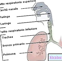
Die Bronchien stellen die an die Trachea angrenzenden Atemwege dar, die sich beim Erwachsenen in Höhe des 4.-5. Primärbronchien wiederum sind in immer kleiner werdende Äste unterteilt, die den sogenannten Bronchialbaum bilden (sie bilden wie eine Pflanze Äste, die immer kleiner werden).

Wie die oberen Atemwege (Nasenhöhlen, Nasopharynx, Pharynx, Larynx und Trachea) sind die Bronchien im Wesentlichen für den Transport von Luft aus der äußeren Umgebung zu den Funktionseinheiten der Lunge, den Alveolen, in denen der Gasaustausch stattfindet (die Lungenbläschen sind kleine luftgefüllte Bläschen, die dicht von Kapillaren umgeben sind und für den Austausch von Sauerstoff und Kohlendioxid verantwortlich sind).
Der Aufbau der Primärbronchien ist identisch mit dem der Trachea; als solche behalten sie eine Knorpelstützstruktur in ihrer Wand bei. Durch die allmähliche Verzweigung in Kanäle geringeren Kalibers entstehen aus den Bronchien die sogenannten Bronchiolen, in denen die oben beschriebene Knorpelstruktur verloren geht.

Nach dem Eindringen in die jeweiligen Lungenlappen wird jeder Lappen- oder Sekundärbronchus in die verschiedenen bronchopulmonalen Segmente unterteilt. In der Lunge verlieren die Lappenbronchien die für die Luftröhre und die Primärbronchien typische knorpelige Stützstruktur (C-Ringe) und werden mit unregelmäßigen Platten aus hyalinem Knorpel bedeckt, während die glatte Muskulatur vollständige Ringe bildet (anders als in der Luftröhre, wo die hinteren Knorpelöffnungen werden vom Trachealmuskel ausgefüllt.) Auf diese Weise haben die intrapulmonalen Bronchien keinen nach hinten abgeflachten Teil mehr, sondern sind vollständig gerundet.

Wenn man den Bronchialbaum betritt, nimmt die Dicke der Bronchialwände zusammen mit dem Kaliber der Atemwege ab, die immer weniger an Knorpelgewebe und zunehmend an Muskelgewebe angereichert sind.
Sobald sie die Lungenlappen durchdringen, teilen sich die Sekundärbronchien in kleinere Äste, die sogenannten Tertiär- (oder Segment-)Bronchien. Jeder von ihnen verzweigt sich, indem er mit kleineren Zweigen unterschiedliche Abschnitte des Lungengewebes bedient, die als bronchopulmonale Segmente bezeichnet werden. Wie in der Abbildung gezeigt, ist jede Lunge tatsächlich durch 10 bronchopulmonale Segmente unterteilt, die durch Bindegewebe voneinander getrennt sind.
Aus den Tertiärbronchien entstehen durch wiederholte Verzweigungen die sogenannten Bronchiolen. Wie erwartet, nimmt auch die Menge an Knorpel in ihrer Wand ab, wenn die Bronchien dünner werden; gleichzeitig nimmt die Zahl der Drüsen und Becherzellen (wichtig zur Verhinderung des Eindringens von Keimen und Staub) ab, während der Beitrag der glatten Muskulatur und des elastischen Gewebes zunimmt und die Epithelhöhe zunehmend abnimmt, während im terminale Bronchiolen die Haarzellen werden quaderförmig (von säulenförmig oder zylindrisch), verlieren die Flimmerhärchen und werden in den für den Gasaustausch verantwortlichen Bereichen weiter abgeflacht (wo "Muskelgewebe fehlt").
Ungefähr 78.000
Die Bronchiolen wiederum teilen sich immer wieder und es entstehen immer kleinere Gänge, die sogenannten terminalen Bronchiolen, mit einem Durchmesser von weniger als 0,5 mm. Diese bilden den letzten Teil des Reizleitungssystems des Atmungssystems, sie versorgen nämlich die Lungenazini mit Luft, wo der Gasaustausch stattfindet.

Die Bronchiolen haben weder Drüsen noch Knorpel in ihrer Wand, während sie mit einer durchgehenden Schicht glatter Muskulatur ausgestattet sind, die die Schleimhaut stützt; sie enthalten auch die sogenannten Clara-Zellen, die die Mucipar-Becherzellen ersetzen und vermutlich dafür verantwortlich sind, das respiratorische Epithel vor Bakterien, Toxinen und Kollaps zu schützen und im Schadensfall für dessen Regeneration zu sorgen.
Untergeordnet setzen sich die terminalen Bronchiolen mit den respiratorischen Bronchiolen fort, die sich von den Vorläufern dadurch erheblich unterscheiden, dass sie mit direkt an ihrer Wand öffnenden Alveolen versehen sind; daher haben sie eine Doppelfunktion, sowohl der Leitung als auch des Gasaustausches.